
"Having this type of technology is very impressive"

"The first time we can really see the spine move ... it’s the technology of the future"

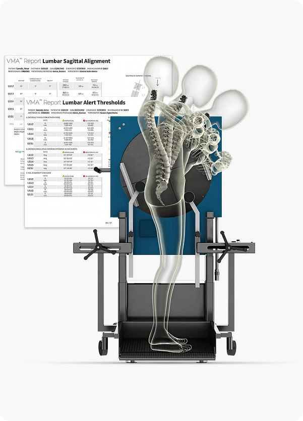
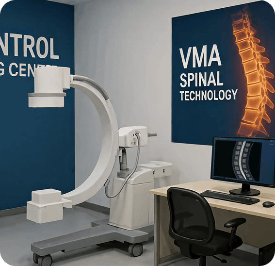
"Too many patients are told their imaging is normal, yet they are still in pain. The VMA gives us objective motion data so we don't miss instability injuries"
"Incorporating VMA in our diagnostic workups has added a new level of objectivity in identifying sites of injury, at the same time as it improves the precision of targeted interventional treatments"


1 Davis RJ, Lee DC, Wade C, Cheng BC. Measurement Performance of a Computer-Assisted Vertebral Motion Analysis System. International Journal of Spine Surgery. 2015.
2 Davis RJ, Lee DC, Wade C, Cheng BC. Variability in Flexion–Extension Radiographs of the Lumbar Spine: A Comparison of Uncontrolled and Controlled Bending. International Journal of Spine Surgery. 2016.
3 Freeman MD, Katz EA, Rosa SL, Gatterman BG, Strömmer EMF, Leith WM. Diagnostic Accuracy of Videofluoroscopy for Symptomatic Cervical Spine Injury Following Whiplash Trauma. International Journal of Environmental Research and Public Health. 2020.
4 Breen A, Muggleton J, Mellor F. An Objective Spinal Motion Imaging Assessment (OSMIA): Reliability, Accuracy, and Exposure Data. BMC Musculoskeletal Disorders. 2006.
5 Steilen D, Hauser R, Woldin B, Sawyer S. Chronic Neck Pain: Making the Connection Between Capsular Ligament Laxity and Cervical Instability. The Open Orthopaedics Journal. 2014.
6 Tominaga Y, Ndu AB, Coe MP, Ivancic PC, Ito S, Rubin W, Panjabi MM. Neck Ligament Strength Is Decreased Following Whiplash Trauma. BMC Musculoskeletal Disorders. 2006.
7 Liu YK, Tipton CM, Matthes RD, Bedford TG, Maynard JA, Carey RA. Healing of Subfailure Ligament Injury: A Comparison Between Immature and Mature Ligaments in a Rat Model. Journal of Orthopaedic Research. 1989.
8 Ivancic PC, Ito S, Tominaga Y, Rubin W, Coe MP, Ndu A, Carlson EJ, Panjabi MM. Whiplash Causes Increased Laxity of Cervical Capsular Ligament. Clinical Biomechanics. 2008.
9 Cholewicki J, McGill S, Wells R, Vernon H. Method for Measuring Vertebral Kinematics From Videofluoroscopy. Clinical Biomechanics. 1991.
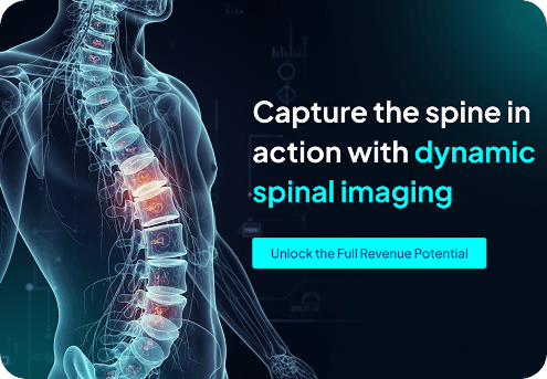
MRI and standard X-ray evaluate the spine at rest often in a non–weight-bearing, supine position. The VMA® analyzes spinal motion while the patient is upright and weight-bearing, capturing how the spine actually functions in daily life. This allows ligament instability to be identified and quantified in a way static or non–weight-bearing imaging cannot reliably achieve.

Yes. The VMA® is designed to integrate into established clinical workflows with minimal disruption. Our team supports setup, reporting, and interpretation so the diagnostic process complements not complicates your practice.
All VMA® studies are independently reviewed and signed by board-certified radiologists. This separation ensures clinical credibility and supports use in treatment planning, insurance review, and med-legal contexts.
The strategy session is a focused, professional discussion to determine clinical fit, diagnostic goals, and operational considerations. It’s designed to clarify whether motion-based diagnosis is appropriate for your practice before any decisions are made.